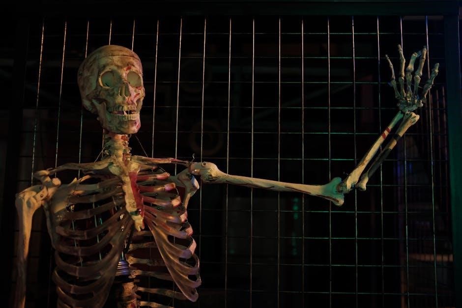
The adult human body consists of 206 bones, varying in size and function, forming the skeletal system’s axial and appendicular divisions. A detailed PDF 206 bones of the body diagram provides a visual guide to understanding their structure and classification, from the smallest ossicles to the largest femur, essential for movement, support, and protection.
Significance of Understanding the Skeletal System
Understanding the skeletal system is crucial for comprehending human anatomy and physiology. It provides insights into how bones support the body, facilitate movement, and protect vital organs. The skeletal system also plays a role in blood cell production and mineral storage. A detailed PDF 206 bones of the body diagram helps visualize bone structure, aiding in education and medical diagnostics. This knowledge is essential for fields like orthopedics and physical therapy, promoting better health and treatment outcomes.
Overview of the 206 Bones Diagram
A comprehensive 206 bones of the body diagram offers a detailed visualization of the human skeletal system. This diagram categorizes bones into the axial (80 bones) and appendicular (126 bones) skeletons. It highlights the structure and arrangement of bones, from the smallest ossicles in the ear to the largest femur. The diagram serves as an essential tool for anatomy students, educators, and healthcare professionals, providing a clear and organized representation of the skeletal framework. It enhances learning and understanding of human anatomy effectively.

The Axial Skeleton
The axial skeleton forms the central axis of the body, comprising 80 bones. It includes the skull, vertebral column, ribs, and sternum, providing structural support and protection for vital organs like the brain and heart.
Overview of the Axial Skeleton
The axial skeleton is the central framework of the body, consisting of 80 bones. It includes the skull, vertebral column, ribs, and sternum. This structure provides essential support for the head, trunk, and internal organs, while also facilitating movement and protection. The axial skeleton forms the foundation for the entire skeletal system, enabling upright posture and housing vital organs. Referencing a PDF 206 bones of the body diagram helps visualize its complex arrangement and functional significance.
Bones of the Skull
The human skull is composed of 22 bones, which fuse together during adulthood. These bones include 8 cranial bones forming the braincase and 14 facial bones shaping the face. The mandible, or jawbone, is the largest facial bone. Together, they protect the brain, house sensory organs, and support facial structures. A detailed PDF 206 bones of the body diagram highlights the skull’s complex anatomy, illustrating how these bones interlock to provide stability and functionality to the head and face.
Vertebral Column and Its Structure

The vertebral column, or backbone, is composed of 33 vertebrae divided into five regions: cervical (7), thoracic (12), lumbar (5), sacrum (5 fused), and coccyx (4 fused). It provides structural support, protects the spinal cord, and facilitates movement. The PDF 206 bones of the body diagram illustrates the vertebrae’s alignment and interconnections, showcasing how they form a flexible yet stable column essential for posture and mobility. Intervertebral discs between vertebrae cushion and absorb shock, maintaining spinal flexibility and overall skeletal integrity.

The Appendicular Skeleton
The appendicular skeleton consists of 126 bones, including the upper and lower limbs, enabling movement and support. A PDF 206 bones of the body diagram illustrates its structure.
Overview of the Appendicular Skeleton
The appendicular skeleton comprises 126 bones, forming the upper and lower limbs, and includes the shoulder and pelvic girdles. It facilitates movement and provides structural support. A PDF 206 bones of the body diagram offers a detailed visualization of this system, showcasing how bones like the humerus, femur, and tibia connect to enable mobility and maintain posture. This framework is essential for locomotion and interacting with the environment, making it a vital part of human anatomy.
Bones of the Upper Limbs
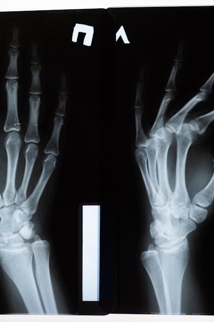
The upper limbs consist of 64 bones, including the scapula, humerus, radius, ulna, carpals, metacarpals, and phalanges. A PDF 206 bones of the body diagram illustrates these bones, showing how they articulate to allow flexible movement. The scapula forms the shoulder, while the humerus connects to the radius and ulna in the forearm. Carpals, metacarpals, and phalanges in the hand enable precise grip and dexterity, making the upper limbs essential for both strength and fine motor skills in daily activities.
Bones of the Lower Limbs
The lower limbs contain 64 bones, including the femur, patella, tibia, fibula, tarsals, metatarsals, and phalanges. These bones provide structural support and enable mobility. The femur, the longest bone, connects to the pelvis, while the tibia and fibula form the shin. Tarsals, metatarsals, and phalanges in the foot allow for balance and locomotion. A PDF 206 bones of the body diagram details their arrangement, showcasing how they contribute to weight-bearing and movement, essential for walking, running, and maintaining posture.

Classification of Bones
Bones are classified into five types: long, short, flat, irregular, and sesamoid. Each category is defined by unique shapes and functions, as detailed in the PDF 206 bones of the body diagram, providing a clear visual guide for understanding their structural differences and roles in the skeletal system.
Long Bones
Long bones are characterized by their greater length compared to their width, with a cylindrical shaft (diaphysis) and rounded ends (epiphyses). Found primarily in the limbs, they include bones like the femur, humerus, tibia, and fibula. These bones provide structural support and leverage for movement, facilitating locomotion and muscle attachment. Their medullary cavities contain bone marrow, essential for blood cell production. The PDF 206 bones of the body diagram highlights their anatomy, showcasing their role in the skeletal system’s functionality and human mobility.
Short Bones
Short bones are cube-shaped or nearly equal in length and width, providing stability and limited movement. Found in the wrists (carpals) and ankles (tarsals), they absorb shock and distribute forces. Their spongy interior supports surrounding structures, while their compact shape allows for flexibility in joints. The PDF 206 bones of the body diagram illustrates their anatomical features, highlighting their role in facilitating movement while maintaining structural integrity in the skeletal system.
Flat Bones
Flat bones are thin, plate-like structures that provide protection and serve as attachment points for muscles. Examples include the skull bones, ribs, sternum, and scapulae. Their broad surfaces allow for muscle attachment, enabling movement and supporting bodily functions. The PDF 206 bones of the body diagram highlights their anatomical details, showcasing how they protect vital organs like the brain and heart while contributing to the skeletal system’s structural integrity and functional diversity.
Irregular Bones
Irregular bones have unique, complex shapes that do not fit into other categories. Examples include vertebrae, pelvic bones, and certain skull bones. These bones protect internal organs, such as the spinal cord, and provide structural support. Their irregular shapes allow for specialized functions, like facilitating movement and absorbing shock. The PDF 206 bones of the body diagram illustrates their intricate structures, helping to identify and understand their roles in the skeletal system’s overall functionality and adaptability.
Sesamoid Bones
Sesamoid bones are small, embedded bones within tendons or muscles, primarily reducing friction and protecting tendons during movement. The patella (kneecap) is the largest sesamoid bone, while others are found in the hands and feet. These bones enhance mechanical advantage, aiding in precise movements. The PDF 206 bones of the body diagram highlights their locations and roles, showcasing their importance in joint stability and muscle function, despite their small size and specialized nature in the skeletal system.
Functions of the Skeletal System
The skeletal system provides support, protects vital organs, facilitates movement, produces blood cells, and stores minerals like calcium and phosphorus, essential for overall bodily function.
Support and Stability
The skeletal system acts as the body’s framework, providing structural support and maintaining posture. Bones such as the spine and pelvis bear the body’s weight, while others like the skull protect vital organs. The arrangement of bones allows for balance and stability, ensuring the body remains upright and functional. This framework is essential for movement and daily activities, enabling the body to withstand external forces effectively; Proper alignment ensures optimal support and prevents potential injuries or deformities.
Facilitation of Movement
The skeletal system plays a crucial role in movement by providing a framework for muscles to attach and act upon. Bones serve as levers, while joints allow for articulation, enabling flexibility and motion. The arrangement of bones and muscles creates a system of leverage, allowing actions like walking, running, and lifting. The skeleton works in harmony with muscles, tendons, and ligaments to facilitate a wide range of movements, from fine motor skills to large-scale mobility, essential for daily activities and maintaining an active lifestyle.
Protection of Internal Organs
The skeletal system serves as a protective framework for vital organs. The skull encases and shields the brain, while the ribcage safeguards the heart, lungs, and liver. Vertebrae protect the spinal cord, and the pelvis encases reproductive organs. Bones act as a barrier, absorbing impacts and preventing damage to delicate internal structures. This protective function ensures the integrity of essential organs, enabling the body to function optimally under various conditions and stresses.
Blood Cell Production
Bones play a crucial role in producing blood cells through hematopoiesis, primarily in the bone marrow of spongy bones like the pelvis, vertebrae, and ribs. Stem cells in the marrow differentiate into red blood cells, white blood cells, and platelets. This process is essential for oxygen transport, immune function, and blood clotting. The bone marrow’s nutrient-rich environment supports continuous blood cell renewal, ensuring the body maintains healthy levels of these vital cells. This function highlights the skeletal system’s role in sustaining life and overall health.
Mineral Storage
Bones serve as a reservoir for essential minerals like calcium and phosphorus, which are vital for maintaining bone strength and overall health. The skeletal system stores approximately 99% of the body’s calcium, which is crucial for nerve and muscle function. When mineral levels in the blood drop, bones release these stored minerals to restore balance. This function ensures proper bodily functions and prevents conditions like osteoporosis, highlighting the skeletal system’s role in mineral homeostasis and overall well-being.
Development of the Skeletal System
The skeletal system begins with 300 bones in infancy, gradually fusing to form 206 bones in adulthood, essential for structure and function.

Bones in Infancy and Childhood
In infancy, the human skeletal system comprises approximately 300 bones, many of which are soft, pliable cartilage. As children grow, these bones gradually ossify and fuse together, a process continuing into early adulthood. This transformation is crucial for developing the structural integrity needed for movement and support. The PDF 206 bones of the body diagram illustrates this progression, showing how the skeletal system matures from a flexible, adaptable structure in infancy to the rigid, 206-bone framework in adulthood. This process ensures optimal growth and functionality.
Bone Growth and Development
Bone growth and development involve the transformation of cartilage into bone tissue through ossification. In childhood, bones are smaller and more flexible, with growth plates facilitating expansion. As individuals mature, these plates gradually close, halting growth. The process ensures bones reach their full size and strength, adapting to the body’s needs. A detailed PDF 206 bones of the body diagram illustrates this journey, highlighting how bones develop and fuse over time to form the adult skeletal system.
Bone Fusion in Adulthood
In adulthood, certain bones fuse together, reducing the total count from the childhood number. This fusion strengthens joints and stabilizes the skeleton. For example, the sacrum and coccyx fuse, and some skull bones unite. A PDF 206 bones of the body diagram illustrates this process, showing how bones mature and solidify, ensuring optimal support and minimizing mobility in key areas, which is crucial for overall skeletal stability and function in adults.

Tools for Studying the Skeletal System
The skeletal system can be studied using PDF diagrams and interactive 3D models. These tools provide detailed visualizations of bone structures, aiding in identification and understanding of the 206 bones.
PDF Diagrams for Visual Learning
PDF diagrams of the 206 bones provide a comprehensive visual guide for anatomy students. These detailed illustrations categorize bones into axial and appendicular skeletons, showcasing their structure and relationships. High-resolution images allow users to zoom in for clarity, making complex anatomical details accessible. The diagrams are downloadable, enabling offline study and reference. They are particularly useful for understanding bone classifications, from long bones like the femur to tiny ossicles in the ear. These resources simplify learning, aiding in the identification and memorization of bone anatomy.
Interactive 3D Models
Interactive 3D models complement PDF diagrams by offering a dynamic way to explore the 206 bones. These models allow users to rotate, zoom, and view bones from multiple angles, enhancing spatial understanding. They are particularly useful for anatomy students to visualize complex bone structures and their relationships. The immersive experience aids in identifying articulations and muscle attachments, making learning engaging and effective. These tools are invaluable for both educational and reference purposes, providing a deeper appreciation of human anatomy.

